Digital Pathology Services
UHN Biospecimen Services will provide consultations, expertise and recommendations for all of your pathology and histology research requirements.
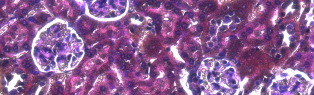
Histology Services
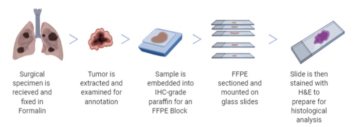
Prior to any histological analysis, biospecimens are prepared to optimizes the longevity, feasibility and retrieval of data down to the cell of each specimen. Utilizing these methods are a way of ensuring our quality assurance to each sample that passes through our facility.
UHN Biospecimen Services offers paraffin services including the processing, embedding, sectioning, and staining of tissues as well as frozen sectioning.
Contact Us
Please contact us to discuss how we can better meet your needs with tissue processing, embedding orientation and staining.
Contact Us
TMA Constructions for Multiplex Histological Analysis
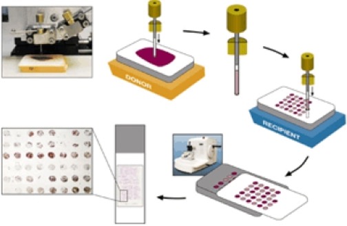
A Tissue Micro Array (TMA) is a unique method by which molecular analysis of tissues is conducted to identify new diagnostic and prognostic markers in oncological disease sites and immunological research.
TMAs are a paraffin embedded block that consist of multiple cylindrical cores of tissue (up to 1,000) that are derived from pre-existing ‘donor’ FFPEs (Formalin Fixed Paraffin Embedded). Guided by H&E slides from the ‘donor’ FFPEs, any morphological area of interest may be selected and cored to be embedded on the TMA. The TMA may then be cut and set for immunohistochemical/immunofluorescent staining, or in situ hybridization for either mRNA or DNA. Tissue sample retrieval is a lengthy process that requires accuracy, precision, and patient consent. Hence, once the tissue is obtained it must be used to its maximum capacity and TMA construction allows for effective usage of tissue. Additionally, TMA construction allows for entire cohort studies to be analyzed in one experimental design in a quick, cost and time efficient manner.
References
- Jawhar N. M. (2009). Tissue Microarray: A rapidly evolving diagnostic and research tool. Annals of Saudi medicine, 29(2), 123–127. https://doi.org/10.4103/0256-4947.51806
- Why use Tissue Microarrays. (2019, September 24). Retrieved June 16, 2020, from https://medicine.yale.edu/pathology/ypts/tissuemicroarrayfacility/why/
Laser Capture Microdissection (LCM) for Molecular Profiling
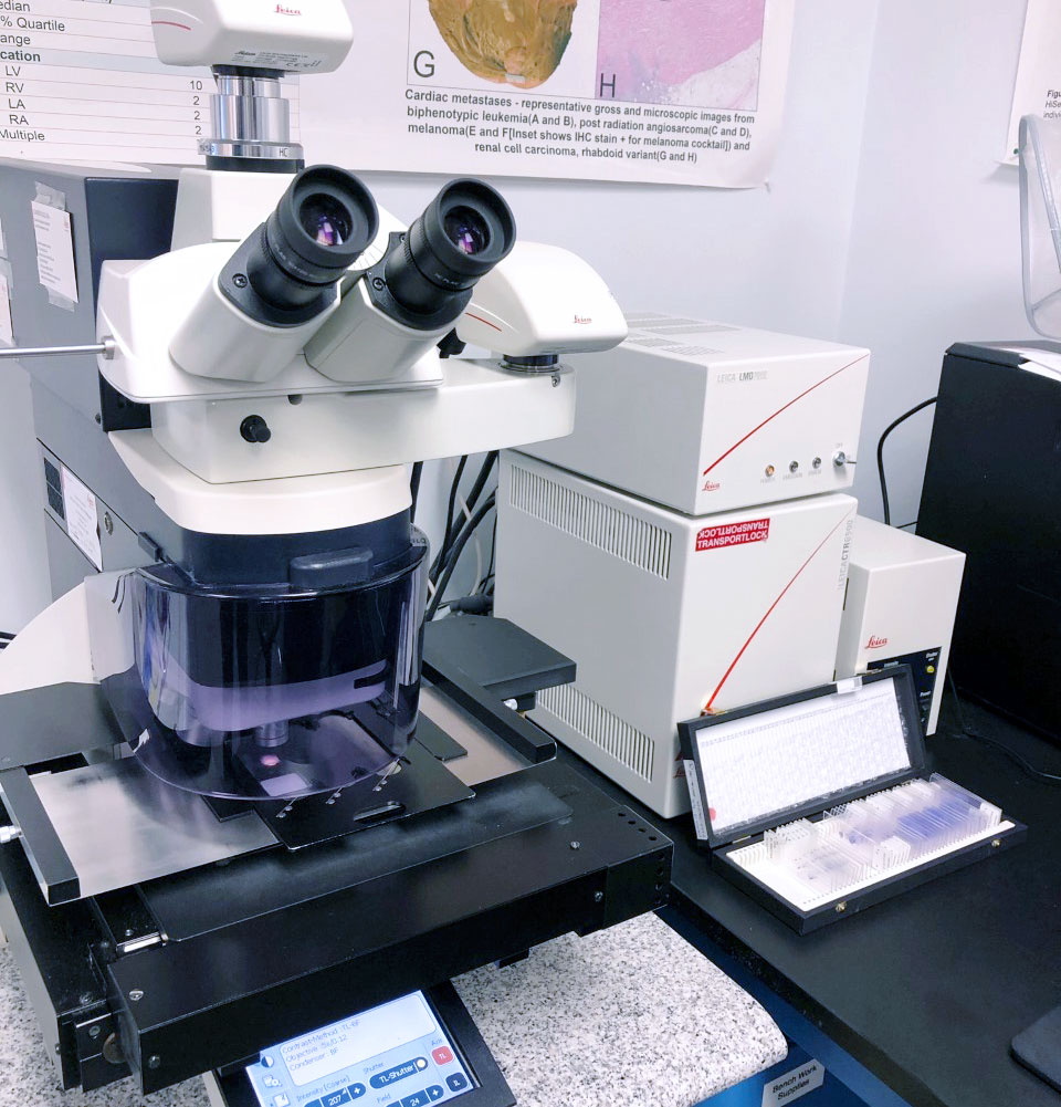
Our Leica LMD6 enables us to provide Laser Capture Microdissection (LCM) of specific disease sites down to the individual cells through contact-free methods minimizing contamination.
It harvests specific cells from a diverse cell population using a precise laser and gravitational force. LCM is an efficient technique that allows for oncological researchers to conduct in-depth analysis on tumour cells and find distinguishing characteristics from non-oncological cells. The optics of the laser grant high precision cuts through tissue in real time from drawing a cut-out of the cellular structures that are required for the specific study group. The cut out is deposited naturally into a capsule providing a high quality, precise and contaminant free sample. These isolated cells are then utilized for DNA, RNA and protein extraction that can be useful for PCR, proteomics analysis, DNA microarray in addition to other analysis.
References:
- Espina, V., Wulfkuhle, J., Calvert, V. et al. Laser-capture microdissection. Nat Protoc 1, 586–603 (2006). https://doi.org/10.1038/nprot.2006.85
- Maloy, S., & Hughes, K. (2013). Brenner’s Encyclopedia of Genetics (2nd ed.). London: Elsevier/Academic Press.
(https://www.sciencedirect.com/topics/medicine-and-dentistry/laser-capture-microdissection)
Slide Scanning
With an enriched history in Biobanking, our team is well equipped with serving your needs to accurately view your biospecimens on a cellular level. Our high volume, automated brightfield scanners (Aperio AT2) precisely digitizes whole slides at optimal quality and a quick turn-around.
Our capture types include:
- 20x
- 40x
- Z-stacking (up to 25 layers)
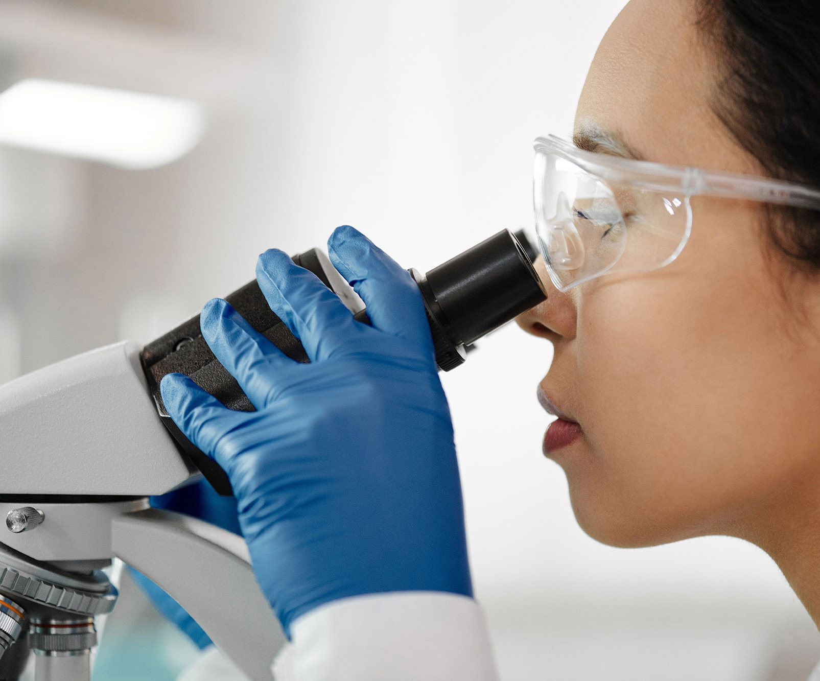
Image Analysis Pathology Annotation
Because modern pathology practice is moving towards a digital and automated workflow, we cater cutting-edge technology to provide you with high-grade histological reviews of your biospecimens.
Working in conjunction with our in-house pathologists and the UHN Pathology Department, our expert pathologists provide a general Pathology Review. Thus, giving initial insight to the histological grade and pathological status of your samples and ensuring the highest quality assurance of your samples and data for your research.
Once the Pathology Review has been cleared, complex data extraction may be performed via machine learning, statistical methods, or other tools for rapid assessments of subcellular components. Data collected may be used for clinical and translational research in oncology and immuno-oncology and extend to other disciplines such as: cardiology, hepatology, gastroenterology, etc.
To view these annotations of your specimens, see our e-library.
E-Library
Welcome to UHN Biospecimen Services E-library!
We offer an e-library of banked biospecimen images in order to facilitate the researcher’s access to a visually available representation through images that will supplement their research.
Coming Soon